

OCCUPATION AND HEALTH ›› 2026, Vol. 42 ›› Issue (20): 2777-2782.
Previous Articles Next Articles
YI Jiping1, YANG Xiuhong1, WANG Yougen2, ZENG Ming3( )
)
Received:2025-08-05
Revised:2025-11-13
Online:2026-10-15
Published:2026-09-14
Contact:
ZENG Ming,E-mail:CLC Number:
YI Jiping, YANG Xiuhong, WANG Yougen, ZENG Ming. Role of the ASMase/Cer signaling pathway in SiO2 inducing NIH3T3 cell fibrosis and the intervention effect of rAC. [J]OCCUPATION AND HEALTH, 2026, 42(20): 2777-2782.
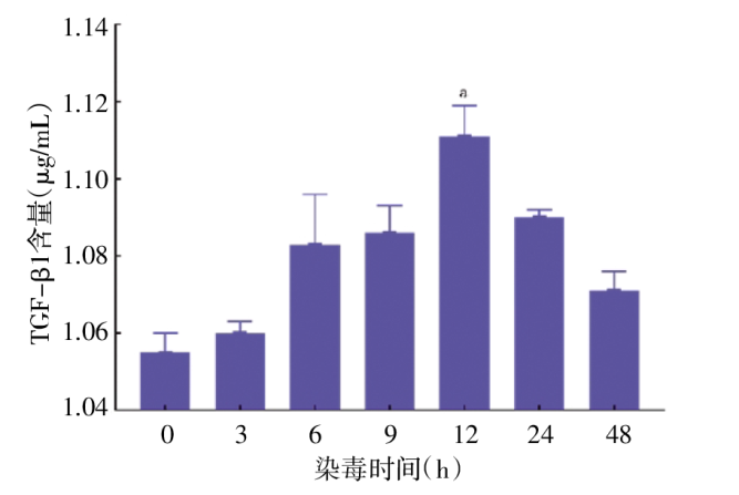
| SiO2剂量(μg/mL) | 细胞存活率(%) | TGF-β1含量(ng/mL) |
|---|---|---|
| 400 | 36.328±0.082a | 1.121±0.008a |
| 200 | 72.949±1.294a | 1.098±0.002a |
| 100 | 73.242±3.981a | 1.086±0.002a |
| 50 | 79.395±0.707a | 1.076±0.016a |
| 25 | 82.617±1.071a | 1.077±0.001a |
| 0 | 100 | 1.051±0.005 |
| SiO2剂量(μg/mL) | 细胞存活率(%) | TGF-β1含量(ng/mL) |
|---|---|---|
| 400 | 36.328±0.082a | 1.121±0.008a |
| 200 | 72.949±1.294a | 1.098±0.002a |
| 100 | 73.242±3.981a | 1.086±0.002a |
| 50 | 79.395±0.707a | 1.076±0.016a |
| 25 | 82.617±1.071a | 1.077±0.001a |
| 0 | 100 | 1.051±0.005 |
| 组别 | ROS(flu-value/mgpro) | MDA(nmol/mgprot) | TGF-β1(ng/mL) |
|---|---|---|---|
| SiO2刺激组 | 51.605±0.769a | 1.250±0.170a | 2.023±0.013a |
| rAC干预组 | 42.830±0.826ab | 0.903±0.098ab | 1.976±0.012ab |
| 对照组 | 37.194±1.017 | 0.694±0.098 | 1.932±0.011 |
| F值 | 63.816 | 9.800 | 34.535 |
| P值 | <0.001 | 0.013 | <0.001 |
| 组别 | ROS(flu-value/mgpro) | MDA(nmol/mgprot) | TGF-β1(ng/mL) |
|---|---|---|---|
| SiO2刺激组 | 51.605±0.769a | 1.250±0.170a | 2.023±0.013a |
| rAC干预组 | 42.830±0.826ab | 0.903±0.098ab | 1.976±0.012ab |
| 对照组 | 37.194±1.017 | 0.694±0.098 | 1.932±0.011 |
| F值 | 63.816 | 9.800 | 34.535 |
| P值 | <0.001 | 0.013 | <0.001 |
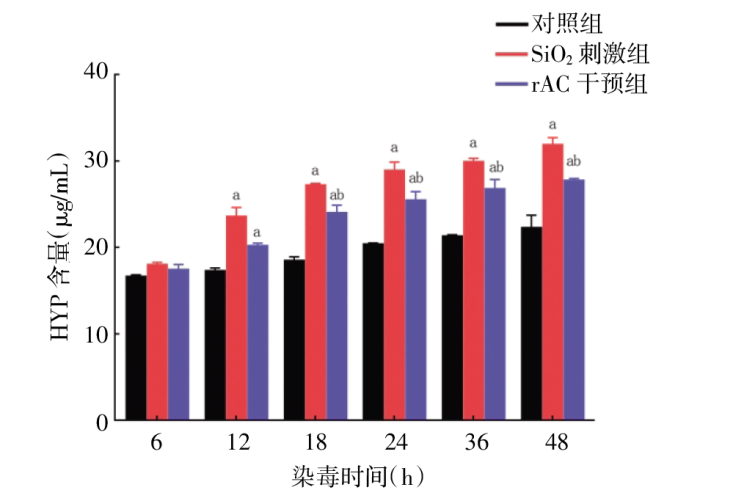
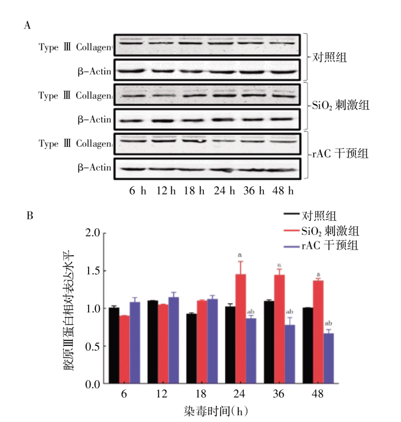
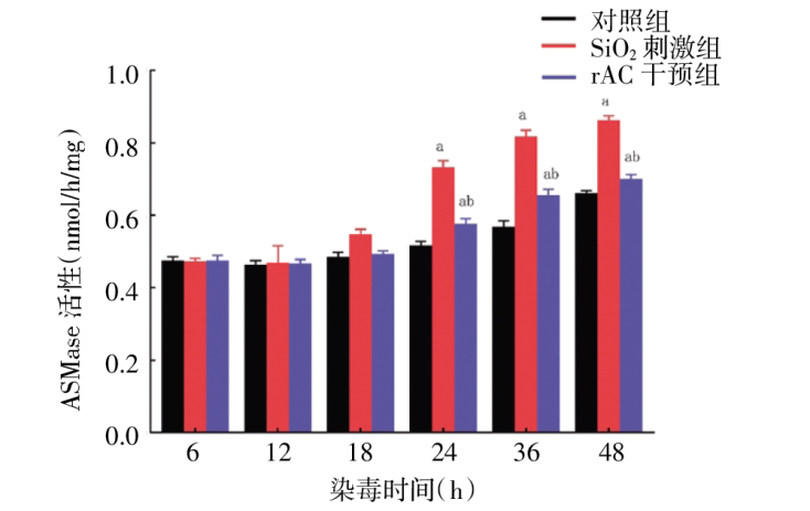
| 指标 | ASMase | Cer | HYP | Col Ⅲ |
|---|---|---|---|---|
| ASMase | 1.000 | |||
| Cer | 0.660b | 1.000 | ||
| HYP | 0.855b | 0.544a | 1.000 | |
| Col Ⅲ | 0.395 | 0.814b | 0.335 | 1.000 |
| 指标 | ASMase | Cer | HYP | Col Ⅲ |
|---|---|---|---|---|
| ASMase | 1.000 | |||
| Cer | 0.660b | 1.000 | ||
| HYP | 0.855b | 0.544a | 1.000 | |
| Col Ⅲ | 0.395 | 0.814b | 0.335 | 1.000 |
| [1] | 国家卫生健康委员会规划发展与信息化司. 2023年我国卫生健康事业发展统计公报[J]. 中国病毒病杂志, 2024, 14(5):416-424. |
| [2] |
GOMEZ-LARRAURI A, PRESA N, DOMINGUEZ-HERRERA A, et al. Role of bioactive sphingolipids in physiology and pathology[J]. Essays Biochem 2020, 64(3):579-589.
DOI URL |
| [3] |
ADAN G A, KARTAL Y M, ISKENDER G, et al. Novel agents targeting bioactive sphingolipids for the treatment of cancer[J]. Curr Med Chem, 2013, 20(1):108-122.
DOI URL |
| [4] |
JAYANT G, KUPERBERG S, SOMNAY K, et al. The role of sphingolipids in regulating vascular permeability in idiopathic pulmonary fibrosis[J]. Biomedicines, 2023, 11(6):1728.
DOI URL |
| [5] |
HUANG F, DU Y, ZHANG X, et al. SiO2 dust induces inflammation and pulmonary fibrosis in rat lungs through activation of ASMase/ceramide pathway[J]. J Appl Toxicol, 2023, 43(9):1319-1331.
DOI URL |
| [6] |
WANG E, HE X, ZENG M, et al. The role of s1p and the related signaling pathway in the development of tissue fibrosis[J]. Front Pharmacol, 2019, 9:1504.
DOI URL |
| [7] | HE X, SCHUCHMAN E H. Potential role of acid sphingomyelinase in environmental health[J]. J Cent South Univ(Med Sci), 2012, 37(2):109-125. |
| [8] |
POCZOBUTT J, EGERSDORF N, HILTABIDLE M S, et al. Short term acid sphingomyelinase deficiency exerts proinflammatory and antiapoptotic effects during LPS-induced lung injury in mice[J]. Am J Respir Cell Mol Biol, 2025, 73(2):275-287.
DOI URL |
| [9] |
LI S, ZHAO J, HAN G, et al. Silicon dioxide-induced endoplasmic reticulum stress of alveolar macrophages and its role on the formation of silicosis fibrosis:A review article[J]. Toxicol Res, 2023, 12(6):1024-1033.
DOI URL |
| [10] |
TANG M, YANG Z, LIU J, et al. Combined intervention with N-acetylcysteine and desipramine alleviated silicosis development by regulating the Nrf2/HO-1 and ASMase/ceramide signaling pathways[J]. Ecotoxicol Environ Saf, 2022, 242:113914.
DOI URL |
| [11] |
MANAGÒ A, BECKER K A, CARPINTEIRO A, et al. Pseudomonas aeruginosa pyocyanin induces neutrophil death via mitochondrial reactive oxygen species and mitochondrial acid sphingomyelinase[J]. Antioxid Redox Signa, 2015, 22(13):1097-1110.
DOI URL |
| [12] |
LIU T T, SUN H F, TANG M Z, et al. Bicyclol attenuates pulmonary fibrosis with silicosis via both canonical and non-canonical TGF-β1 signaling pathways[J]. J Transl Med, 2024, 22(1):1-19.
DOI |
| [13] | BONFIELD T L. Membrane lipids and CFTR:The Yin/Yang of efficient ceramide metabolism[J]. J Transl Med, 2020, 202(8):1074-1075. |
| [14] | BECKER K A, TÜMMLER B, GULBINS E, et al. Accumulation of ceramide in the trachea and intestine of cystic fibrosis mice causes inflammation and cell death[J]. Am J Respir Crit Care Med, 2010, 403(3):368-374. |
| [15] | BECKER K A, RIETHMÜLLER J, ZHANG Y, et al. The role of sphingolipids and ceramide in pulmonary inflammation in cystic fibrosis[J]. Open Respir Med J, 2010, 4(2):39-47. |
| [16] |
SUNG E A, DOZMOROV M G, SONG S, et al. Ablation of LRP6 in alpha-smooth muscle actin-expressing cells abrogates lung inflammation and fibrosis upon bleomycin-induced lung injury[J]. FEBS Letters, 2025, 599(10):1468-1480.
DOI URL |
| [17] |
ZIOBRO R, HENRY B, EDWARDS M J, et al. Ceramide mediates lung fibrosis in cystic fibrosis[J]. Biochem Biophys Res Commun, 2013, 434(4):705-709.
DOI URL |
| [18] |
HE X, OKINO N, DHAMI R, et al. Purification and characterization of recombinant,human acid ceramidase,Catalytic reactions and interactions with acid sphingomyelinase[J]. J Biol Chem, 2003, 278(35):32978-32986.
DOI URL |
| [19] |
SIMONARO C M, SACHOT S, GE Y, et al. Acid ceramidase maintains the chondrogenic phenotype of expanded primary chondrocytes and improves the chondrogenic differentiation of bone marrow-derived mesenchymal stem cells[J]. PLoS One, 2013, 8(4):e62715.
DOI URL |
| [20] |
XU S W, LIU S, EASTWOOD M, et al. Rac inhibition reverses the phenotype of fibrotic fibroblasts[J]. PLoS One, 2009, 4(10):e7438.
DOI URL |
| [21] | BECKER K A, VERHAEGH R, VERHASSELT H L, et al. Acid ceramidase rescues cystic fibrosis mice from pulmonary infections[J]. Infect Immun, 2021, 89(2):e00677-20. |
| [22] |
LIU S, KAPOOR M, LEASK A. Rac1 expression by fibroblasts is required for tissue repair in vivo[J]. Am J Pathol, 2009, 174(5):1847-1856.
DOI PMID |
| Viewed | ||||||
|
Full text |
|
|||||
|
Abstract |
|
|||||
 津公网安备12010202000831号 津ICP备11005889号-6